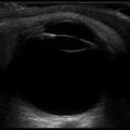
Ultrasonography
Ultrasonography the imaging of deep structures of the body by recording the echoes of pulses of ultrasonic waves directed into the tissues and reflected by tissue planes where there is a change in density.
An eye and orbit ultrasound is a test to look at the eye area, and to measure the size and structures of the eye.
Why the Test is Performed
The ultrasound can examine the farthest part of the eyeball when you have cataracts or other conditions that make it hard for the doctor to look into your eye. The test may help diagnose retinal detachment or other disorders when the eye is not clear and the ophthalmologist cannot use routine examining equipment.
How the Test is Performed
The test is usually done by experts.
You usually sit in a chair. Your eye is numbed with medicine (anesthetic drops). The ultrasound wand (transducer) is placed against the front surface of the eye.
The ultrasound uses high-frequency sound waves that travel through the eye. Reflections (echoes) of the sound waves form a picture of the structure of the eye. The test takes about 15 minutes.


 3i Global Inc.