Page 3
Page 2
Page 1
Laser Treatments
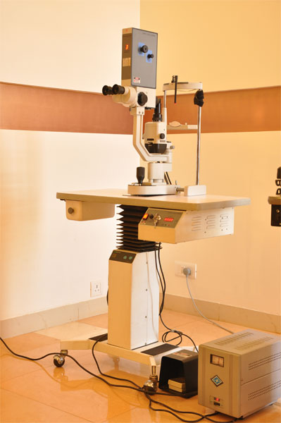
Green Laser Treatment
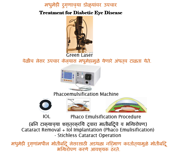
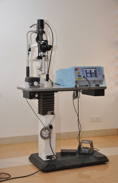
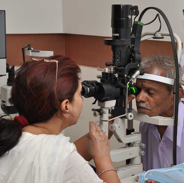
Green laser photocoagulator is a laser-based medical equipment extensively used for the treatment of diabetic retinopathy. Diabetic retinopathy is a disease which causes the growth of excess fragile blood vessels in the retina of the patient suffering from prolonged diabetes. The rupture of these blood vessels causes bleeding and subsequently leads to blindness. The green light from the photocoagulator can reach the retina without much absorption at the intermediate ocular media and gets absorbed at the retina, enabling controlled cutting and blocking of the excess blood vessels leading to simple and less painful treatment of the disease with faster healing than the conventional surgical methods.
It is the most effective and safest laser with the property of maximum absorption by hemoglobin and melanin and least absorption by xanthophylls pigment of macula.
DCR Laser Treatment
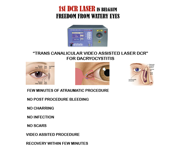
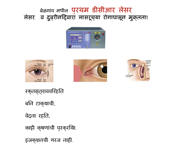
Dacryocystorhinostomy (DCR) is a surgical procedure to restore the flow of tears into the nose from the lacrimal sac when the nasolacrimal duct does not function.Endoscopic laser dacryocystorhinostomy (DCR) enables an obstructed lacrimal sac to be opened through an intranasal approach, avoiding the need for a skin incision.
The operation can also be performed endoscopically through the nose where an opening is fashioned in the lacrimal sac from within the nose. Newer techniques and advances in endoscopic surgery has increasingly made this approach the procedure of choice. The advantages include lesser peri-operative morbidity, no scar and a high success rate. Even revision surgery done by the endoscopic route is easier.
With the advent of nasal endoscopes endoscopic dacryocystorhinostomy is becoming popular. In this procedure a nasal endoscope is used to visualise the lacrimal sac through the nasal cavity. The bone covering the lacrimal sac is nibbled out. The medial wall of the sac is excised facilitating drainage of tears into the nasal cavity. This procedure avoids scar.
The endoscopic approach has several advantages, including the following:
- It provides a better aesthetic result with no external scar.
- It allows a one-stage procedure to also correct associated nasal pathology that may be causative.
- It avoids injury to the medial canthus and/or pathologic scar formation.
- It is much less bloody and messy than the external approach.
- The success rate is comparable to the external approach.
Computerised Perimetry
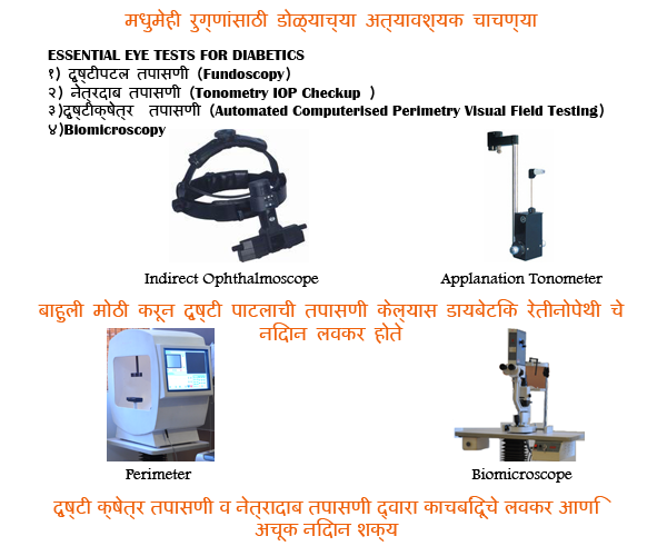
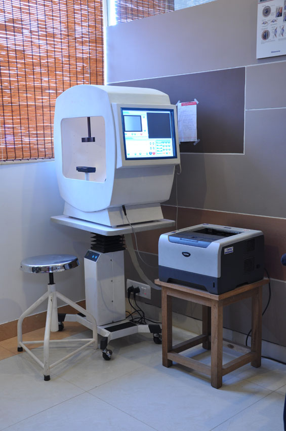 During the last few years computerized perimetry has become a clinical reality. This new technique eliminates the operator’s error, ensures reproducibility of test procedures and parameters and makes visual field testing of large numbers of patients possible. Great differences exist between computerized perimeters. Differences in hardware for example, the way in which stimuli or field charts are produced may be striking but even more important are differences in software, especially test algorithms. The results obtained with some instruments and test programmes are of such high quality that the same level of performance may be almost impossible to obtain with manual perimetry, at least in a clinical setting. Nevertheless, improvements can be expected in the future particularly in adaptive tests and in computerized interpretation of the fields.
A visual field test is an examination that may be performed to analyze a patient’s visual field. The exam may be performed by a technician in one of several ways. The test may be performed by a technician directly, with the assistance of a machine, or completely by an automated machine. Machine based tests aid diagnostics by allowing a detailed printout of the patient’s visual field.
Computerized perimeters have increased the precision with which visual fields may be tested. The purpose here is to present considerations for properly performing automated perimetry. Selecting the correct perimeter to complement specific practices is discussed, and various test strategies available on automated perimeters are reviewed. Selection of the appropriate program to provide the clinician with the information needed is essential. A step-by-step discussion as to proper performance of automated perimetry includes factors that should be considered so that consistency between visual field examinations can be maintained. Confounding variables, such as the learning effect, pupil size, and various ocular pathologies, are discussed with respect to their effects on automated perimetric results.
Ultrasonography
Ultrasonography the imaging of deep structures of the body by recording the echoes of pulses of ultrasonic waves directed into the tissues and reflected by tissue planes where there is a change in density.
An eye and orbit ultrasound is a test to look at the eye area, and to measure the size and structures of the eye.
Why the Test is Performed
The ultrasound can examine the farthest part of the eyeball when you have cataracts or other conditions that make it hard for the doctor to look into your eye. The test may help diagnose retinal detachment or other disorders when the eye is not clear and the ophthalmologist cannot use routine examining equipment.
How the Test is Performed
The test is usually done by experts.
You usually sit in a chair. Your eye is numbed with medicine (anesthetic drops). The ultrasound wand (transducer) is placed against the front surface of the eye.
The ultrasound uses high-frequency sound waves that travel through the eye. Reflections (echoes) of the sound waves form a picture of the structure of the eye. The test takes about 15 minutes.
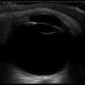
Normal Eye
Biometry
Biometry, or Biometrics, is a division of Biology that uses statistical applications for conducting the study of living things. Biometric tools are widely used in studying growth, biological similarities and differences, authentication of individuals based on his/ her behavioral and physiological characteristics.
The measurement of the various dimensions of the eye and of its components and their interrelationships. The axial length and the corneal curvature are essential measurements to predict the correct lens power of an intraocular lens. There are several biometers which are used prior to cataract surgery some based on ultrasound, others on optical systems. A commonly used optical biometry method called partial coherence interferometry (PCI) (e.g. uses infrared laser light and provides a measurement of axial length, lens thickness, anterior chamber depth and corneal curvature). It also includes software for the calculation of an intraocular lens power using a selection of formulae. It is not appropriate for eyes with dense cataracts or severe corneal oedema, in which case ultrasonography is preferable
A-scan is an amplitude modulation scan. It gives the information in the form of one dimensional. it is used to detect the presence of flaws in the materials. A-scan ultrasound biometry, commonly referred to as an A-scan, is routine type of diagnostic test used in ophthalmology. The A-scan provides data on the length of the eye, which is a major determinant in common sight disorders. The most common use of the A-scan is to determine eye length for calculation of intraocular lens power. Briefly, the total refractive power of the emmetropic eye is approximately 60. Of this power, the cornea provides roughly 40 diopters, and the crystalline lens 20 diopters. When a cataract is removed, the lens is replaced by an artificial lens implant. By measuring both the length of the eye (A-scan) and the power of the cornea (keratometry), a simple formula can be used to calculate the power of the intraocular lens needed. There are several different formulas that can be used depending on the actual characteristics of the eye.
The other major use of the A-scan is to determine the size and ultrasound characteristics of masses in the eye, in order to determine the type of mass. This is often termed quantitative A-scan.







 3i Global Inc.